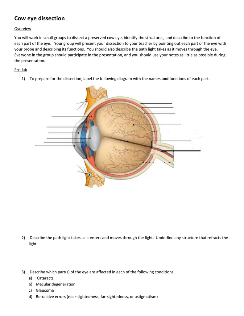
44 eye diagram with labels and functions
PDF Parts of the Eye - National Eye Institute | National Eye Institute To understand eye problems, it helps to know the different parts that make up the eye and the functions of these parts. Here are descriptions of some of the main parts of the eye: ... Handout illustrating parts of the eye Keywords: parts of the eye, eye diagram, vitreous gel, iris, cornea, pupil, lens, optic nerve, macula, retina ... eye diagram labeled brain function structure functions diagram lobes macmillan labelled showing. Cow Eye Dissection - YouTube . cow eye dissection. Label The Muscles Of The Eye - PurposeGames . purposegames. 3d Eye Model 32 Pcs Assembled Human Anatomy Model New 3D Structure Of . auge. Photoreceptor Cell ...
The Eyes (Human Anatomy): Diagram, Optic Nerve, Iris, Cornea ... - WebMD Just behind the iris and pupil lies the lens, which helps focus light on the back of your eye. Most of the eye is filled with a clear gel called the vitreous. Light projects through your pupil and...
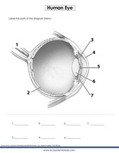
Eye diagram with labels and functions
Anatomy of the eye: Quizzes and diagrams - Kenhub Take a look at the diagram of the eyeball above. Here you can see all of the main structures in this area. Spend some time reviewing the name and location of each one, then try to label the eye yourself - without peeking! - using the eye diagram (blank) below. Unlabeled diagram of the eye. Click below to download our free unlabeled diagram of ... Liver Diagram with Detailed Illustrations and Clear Labels Liver – Anatomy, Functions, And Liver Diseases; Also Read: 6 Facts Everyone Should Know About The Liver; Fatty Liver Symptoms – Explore The Signs, Indications And Causes; 12 Alarming Symptoms of Liver Problems You Shouldn’t Ignore; A Brief Account Of Hepatic Portal System And Its Significance; Human Body – Anatomy and Physiology of ... Eye anatomy: A closer look at the parts of the eye The iris of the eye functions like the diaphragm of a camera, controlling the amount of light reaching the back of the eye by automatically adjusting the size of the pupil (aperture). The eye's crystalline lens is located directly behind the pupil and further focuses light.
Eye diagram with labels and functions. Label Parts of the Human Eye - University of Dayton Label Parts of the Human Eye. Select One Anterior Chamber Ciliary Body Cornea Fibrous Tunic Iris Lateral Rectus Muscle Lens Medial Rectus Muscle Optic Disk Optic Nerve Pupil Retina Vascular Tunic Vitreous Nerve. Select One Anterior Chamber Ciliary Body Cornea Fibrous Tunic Iris Lateral Rectus Muscle Lens Medial Rectus Muscle Optic Disk Optic ... Parallel categories diagram in Python - Plotly Multi-Color Parallel Categories Diagram¶. The color of the ribbons can be specified with the line.color property. Similar to other trace types, this property may be set to an array of numbers, which are then mapped to colors according to the the colorscale specified in the line.colorscale property.. Here is an example of visualizing the survival rate of passengers in the titanic … Label the microscope — Science Learning Hub Jun 08, 2018 · Labels. Description. eye piece lens. The lens you look through – normally 10x or 15x magnification. coarse focus adjustment. Moves the lens up or down and adjusts focus. fine focus adjustment. Moves the lens in order to make very small adjustments to gain better focus. base. The bottom of the microscope used for stability. high-power objective Labelled Diagram of Human Eye, Explanation and Function - VEDANTU The human eye is a part of the sensory nervous system. Labeled Diagram of Human Eye The eyes of all mammals consist of a non-image-forming photosensitive ganglion within the retina which receives light, adjusts the dimensions of the pupil, regulates the availability of melatonin hormones, and also entertains the body clock.
Diagram of the Eye - Home - Lions Eye Institute In order for the eye to work at its best, all parts must work well collectively. To understand the eye and its functions, it's important to understand how the eye works, see below diagrams for both the external eye and the internal eye. The External Eye Instructions Click the parts of the eye to see a description for each. Labelling the eye — Science Learning Hub In this interactive, you can label parts of the human eye. Use your mouse or finger to hover over a box to highlight the part to be named. Drag and drop the text labels onto the boxes next to the eye diagram If you want to redo an answer, click on the box and the answer will go back to the top so you can move it to another box. BYJUS BYJUS The Human Eye - Diagram, Parts, Working, Function and Work of ... - VEDANTU The human eye operates similar to a digital camera in several ways: Light focuses mainly on the cornea, which acts like a camera lens. The iris controls the light that reaches the eye by adjusting the size of the pupil, and thus it functions like the diaphragm of a camera. The lens of the eye is located behind the pupil, and it focuses light.
Eye Anatomy: Parts of the Eye and How We See Here is a tour of the eye starting from the outside, going in through the front and working to the back. Eye Anatomy: Parts of the Eye Outside the Eyeball The eye sits in a protective bony socket called the orbit. Six extraocular muscles in the orbit are attached to the eye. These muscles move the eye up and down, side to side, and rotate the eye. Your Eyes (for Kids) - Nemours KidsHealth It is a very important part of the eye, but you can hardly see it because it's made of clear tissue. Like clear glass, the cornea gives your eye a clear window to view the world through. Iris Is The Colorful Part. Behind the cornea are the iris, the pupil, and the anterior chamber. The iris (say: EYE-riss) is the colorful part of the eye. When ... Create a Briliant Process Flow Diagram with Canva There are lots of ways to use color in a process flow diagram. You could have all the arrows in one part of the process the same color to make it clear they relate to that process. For example, you could use colors like blue and green to represent a cooling process or red and yellow to represent something being heated. To recolor any element or text in your design, select it, then … Label the microscope — Science Learning Hub 08/06/2018 · All microscopes share features in common. In this interactive, you can label the different parts of a microscope. Use this with the Microscope parts activity to help students identify and label the main parts of a microscope and then describe their functions.. Drag and drop the text labels onto the microscope diagram. If you want to redo an answer, click on the box and …
31 Label The Eye Quiz - Best Labeling Ideas
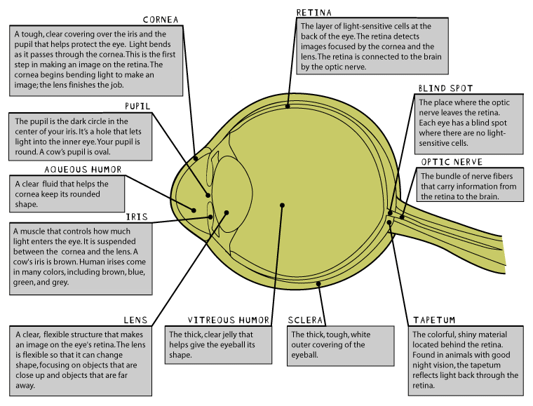
Human Eye Diagram, How The Eye Work -15 Amazing Facts of Eye First, light rays enter the eye through the cornea, the clear front "window" of the eye. The dome shaped cornea bends light to help the eye focus. From the cornea, the light passes through an opening called the pupil. The amount of light passing through is controlled by the iris, or the colored part of your eye.

Male Anatomy Diagram Unlabeled : Sc 912 L 16 13 Reproductive System - Blank ear diagram human ...
Eye anatomy: Muscles, arteries, nerves and lacrimal gland - Kenhub Bony cavity within the skull that houses the eye and its associated structures (muscles of the eye, eyelid, periorbital fat, lacrimal apparatus) Bones of the orbit. Maxilla, zygomatic bone, frontal bone, ethmoid bone, lacrimal bone, sphenoid bone and palatine bone. Structure of the eye. Cornea, anterior chamber, lens, vitreous chamber and ...
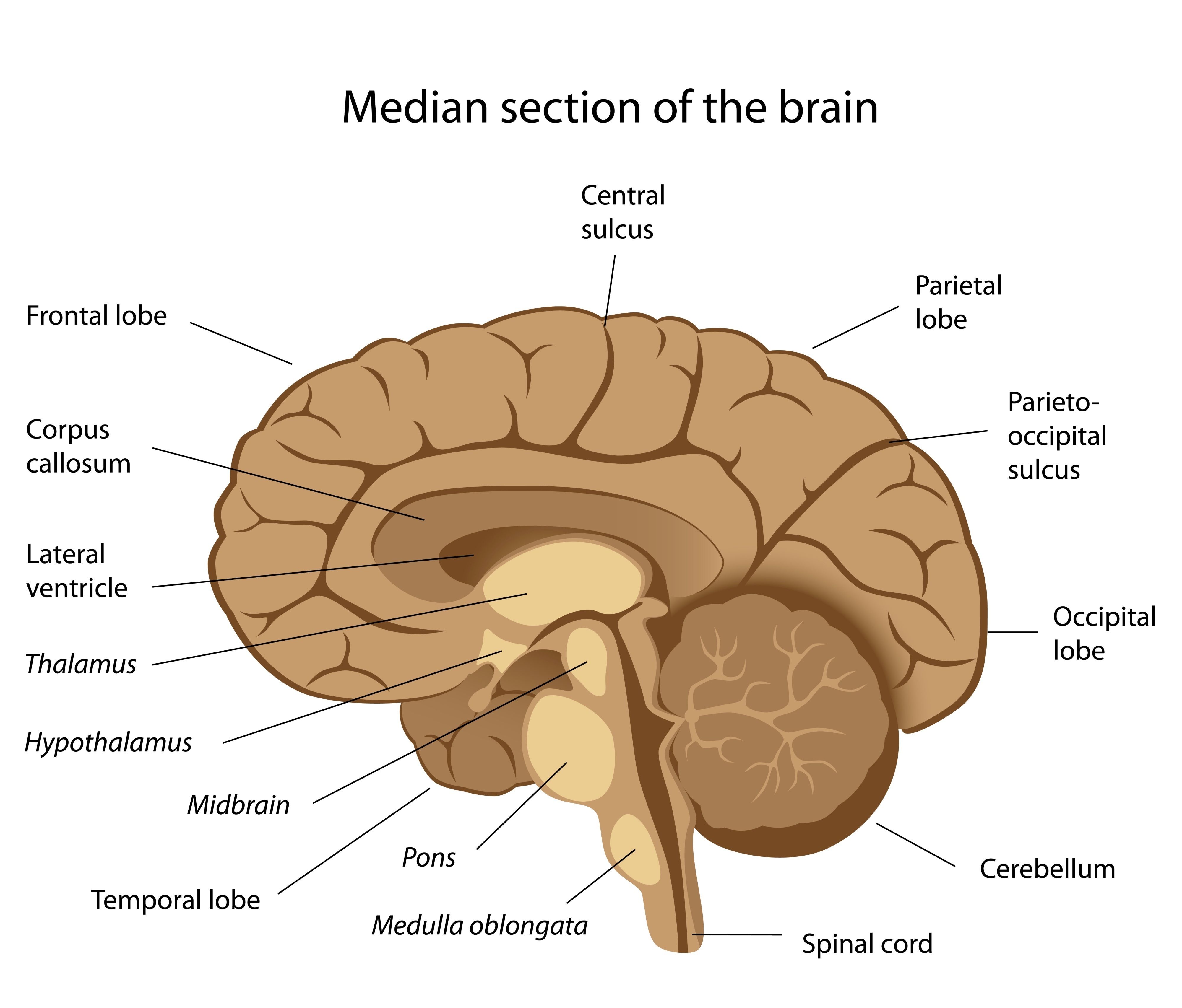
THE BRAIN FROM TOP TO BOTTOM
Eye Anatomy: 16 Parts of the Eye & Their Functions The following are parts of the human eyes and their functions: 1. Conjunctiva The conjunctiva is the membrane covering the sclera (white portion of your eye). The conjunctiva also covers the interior of your eyelids. Conjunctivitis, often known as pink eye, occurs when this thin membrane becomes inflamed or swollen.

The brain - structure and function - Cancer Information - Macmillan Cancer Support
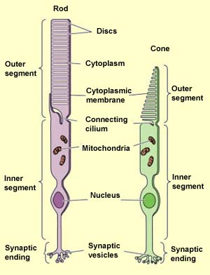
Eye Anatomy Diagram - EnchantedLearning.com Retina - light-sensitive tissue that lines the back of the eye. It contains millions of photoreceptors (rods and cones) that convert light rays into electrical impulses that are relayed to the brain via the optic nerve. Rods - cells the in the retina that sense brightness (they are photoreceptors). Night vision involves mostly rods (not cones).

Module 1: Labeled Diagram of the Eye | Eye health | Pinterest | Activities
Eye Diagram Teaching Resources | Teachers Pay Teachers The Human Eye Overview Reading Comprehension and Diagram Worksheet. by. Teaching to the Middle. 63. $1.50. Zip. This passage briefly describes the human eye (900-1000 Lexile). 14 questions (matching and multiple choice) assess students' understanding. Students label a diagram of 6 parts of the eye. I've included a color and BW version, as well ...
The Eye Diagram Label Worksheets (Differentiated) by zmzb | Teaching Resources
Parts Of The Eye Labeled Diagram Model And Their Function Parts of the eye-labeled diagram model are divided into three groups: the external outer layer, the middle layer, and the inner back layer. The outer layer is responsible for protecting the eye from environmental toxins and debris. The middle layer includes cells that allow light to enter and travel through the back layer to the retina.

draw a diagram of the human eye as seen in a vertical section and label the part which suits the ...
Eye Anatomy | Definition, Structure & Functions - iBiologia Diagram of Human Eye with Labelling. Eye Anatomy Complete Physiology of Eye is described below in the given paragraph: The eye is rather like a living Camera. Each eye is a liquid-filled ball 2.5 cm in diameter. At the front of the eye is a clear, round window called the cornea. Behind the cornea is a "lens.

May I have a simplest diagram of an eye, Please - Science - The Human Eye and the Colourful ...
Label the Eye Worksheet – Teacher-Made Learning Resources In this resource, you’ll find a 2-page PDF that is easy to download, print out, and use immediately with your class. The first page is a labelling exercise with two diagrams of the human eye. One is a view from the outside, and the other is a more detailed cross-section. Challenge learners to label the parts of the eye diagram. On the second page, you’ll find a set of answers …

Blank Eye Diagram - Cliparts.co
The Eye Diagram: What is it and why is it used? The eye diagram is used primarily to look at digital signals for the purpose of recognizing the effects of distortion and finding its source. To demonstrate using a Tektronix MDO3104 oscilloscope, we connect the AFG output on the back panel to an analog input channel on the front panel and press AFG so a sine wave displays. Then we press Acquire.
Labeled Eye Diagram - ClipArt Best
Parallel categories diagram in Python - Plotly Basic Parallel Categories Diagram with graph_objects¶ This example illustrates the hair color, eye color, and sex of a sample of 8 people. The dimension labels can be dragged horizontally to reorder the dimensions and the category rectangles can be dragged vertically to reorder the categories within a dimension.
Free Brain Diagram, Download Free Brain Diagram png images, Free ClipArts on Clipart Library
Consumer Updates | FDA The site is secure. The https:// ensures that you are connecting to the official website and that any information you provide is encrypted and transmitted securely.

Structure and functions of the eye by sgreen2 - Teaching Resources - Tes
Eye Diagram With Labels and detailed description - BYJUS A brief description of the eye along with a well-labelled diagram is given below for reference. Well-Labelled Diagram of Eye The anterior chamber of the eye is the space between the cornea and the iris and is filled with a lubricating fluid, aqueous humour. The vascular layer of the eye, known as the choroid contains the connective tissue.
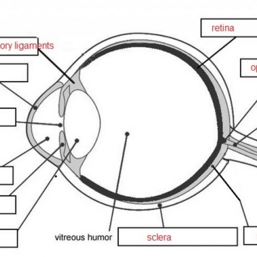
Labeled Eye Diagram - ClipArt Best
Generate eye diagram - MATLAB eyediagram - MathWorks eyediagram(x,n) generates an eye diagram for signal x, plotting n samples in each trace. The labels on the horizontal axis of the diagram range between –1/2 and 1/2. The function assumes that the first value of the signal and every nth value thereafter, occur at integer times.

Blank Ear Diagram | Human ear diagram, Ear anatomy, Ear diagram
Liver Diagram with Detailed Illustrations and Clear Labels Liver – Anatomy, Functions, And Liver Diseases; Also Read: 6 Facts Everyone Should Know About The Liver; Fatty Liver Symptoms – Explore The Signs, Indications And Causes; 12 Alarming Symptoms of Liver Problems You Shouldn’t Ignore; A Brief Account Of Hepatic Portal System And Its Significance; Human Body – Anatomy and Physiology of ...

Diagram of the Ear
Labelling the eye — Science Learning Hub Labelling the eye Add to collection The human eye contains structures that allow it to perceive light, movement and colour differences. In this activity, students use online or paper resources to identity and label the main parts of the human eye. By the end of this activity, students should be able to: identify the main parts of the human eye
Eye Diagram With Labels And Functions - Aflam-Neeeak
Control Unit Installation and Operation Guide Please Read between any Eye QS control unit and any other power supply, including another GRAFIK Eye QS control unit. Refer to the QS Link Power Draw Units specification submittal (Lutron P/N 369405) for more information concerning PDUs. 1234 12 ABC 123456LN Example: Emergency lighting interface (maximum 1) Note: The GRAFIK Eye QS control unit
Post a Comment for "44 eye diagram with labels and functions"